|
NOVIDADES
Stanford chemists have developed a new deep-tissue imaging technique that can see beneath the skin of living subjects to illuminate buried tumors with unparalleled clarity. In a new study published in the journal Nature Biotechnology ("In vivo molecular imaging for immunotherapy using ultra-bright near-infrared-IIb rare-earth nanoparticles"), the researchers demonstrate how their technique can be used to predict the response of cancer patients to immunotherapy and to track their progress following treatment. 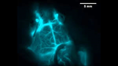 Credits: Stanford University
“We call this infrared vision for non-invasively peering into biological tissues,” said study leader Hongjie Dai, the J.G. Jackson and C.J. Wood Professor in Chemistry in Stanford’s School of Humanities and Sciences. The technique relies on nanoparticles containing the element erbium, which belongs to a class of so-called rare-earth minerals prized by chemists for their unique ability to glow in the infrared. The team covered the nanoparticles in a chemically engineered coating that helps the particles dissolve in the bloodstream and also makes them less toxic and exit the body quicker. In addition, the coating provides anchoring points for molecules that that act like guided missiles to locate and attach to specific proteins on cells. In a video highlighting the power of their nanoprobes, the branching blood vessels of a brain inside a living mouse are limned in a teal light and shift as the creature moves its head. “Our approach allows for seeing into an intact mouse brain while conventional approaches see only the scalp,” said study co-first author Zhuoran Ma, a graduate student in Dai’s lab. In the study, the researchers show their technique can identify tumors in mice carrying a protein that makes them vulnerable to cancer drugs that activate the body’s own immune system. This approach could provide a noninvasive way of identifying patients who would respond well to those drugs without needing to take a sample, or biopsy, of the tumor, as they currently need to do. Similarly, erbium nanoparticles can monitor patients’ responses following cancer therapy to track whether they are responding to the drugs and if their tumors have shrunk. As a demonstration, Dai and his colleagues combined their erbium nanoparticles with another infrared imaging technique to simultaneously image two molecular targets –cancer cells and T-cells – simultaneously. The result is a layered, multi-colored snapshot showing T-cells homing in on a tumor from elsewhere in the mouse’s body. The researchers said their technique could also enable surgeons to more precisely excise tumors and aid biologists and medical researchers in studying fundamental processes within cells. By Ker Than, Stanford University. Accessed: Oct 08, 2019. |
|||||||||||||||||||||||||