|
NOVIDADES
In an incredibly fast piece of research, scientists from the University of Texas at Austin and the National Institutes of Health have released a cryo-electron microscopy (cryoEM) structure of part of SARS-CoV-2, the novel coronavirus that has infected tens of thousands of people and killed more than 2,000 since the end of December (Science 2020 DOI: 10.1126/science.abb2507). 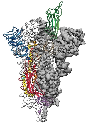 Side view of the SARS-CoV-2 spike protein with one receptor binding domain shown in colored ribbons. Credit: Jason McLellan/University of Texas at Austin
UT Austin’s Jason McLellan and his colleagues have spent many years studying other coronaviruses and had already figured out how to use select mutations to lock coronavirus spike proteins into a shape that is conducive for structural studies. After they got the genome sequence of the virus, it took the team just two weeks to design and produce samples of the stabilized spike protein. After collecting data on their stabilized spike protein samples using a cryo-electron microscope, the researchers spent 12 days reconstructing the 3-D structure. They published the results on bioRXiv on Feb. 15, and the paper was rushed through peer review before being published by Science on Feb. 19 (DOI: 10.1126/science.abb2507). C&EN has made this story and all of its coverage of the coronavirus epidemic freely available during the outbreak to keep the public informed. To support our journalism, become a member of ACS or sign up for C&EN's weekly newsletter. “This is stunning work, illustrating the power of molecular biology in combination with cryoEM,” says Alice Clark, a structural biologist at the University of Wolverhampton. “How quickly this work was possible is a credit to both the scientists involved in this structure, and the recent advances in cryoEM as a technique.” By Laura Howes. Chemical and Engineering News. Posted: Feb. 19, 2020.
An unobstructed view into the cell.
A potential difference for single-particle cryo-EM. The cryo-EM revolution: fueling the next phase.
|
|||||||||||||||||||||||||